ไทย
ไทย- ×
 High Blood Sugar Package 1 × ฿2,790
 High Blood Sugar Package
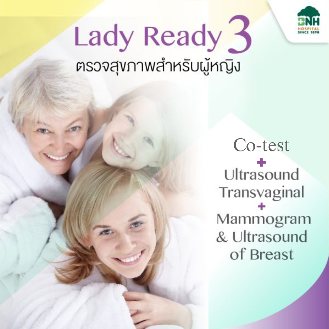
Lady Ready 3 :Cotest (Pap smear and HPV DNA test) + Transvaginal Ultrasound + Mammogram with ultrasound of breast
Original price was: ฿14,900.฿10,000Current price is: ฿10,000.
Package Includes
- Cervical cancer screening with Co-Test(Pap smear and HPV DNA test)
- Vaginal ultrasound to determine the integrity of the uterus and ovaries.
- Breast cancer screening using mammogram and breast ultrasound.
*This price includes doctor fees and hospital service fees.
Lady Ready 3 :Cotest (Pap smear and HPV DNA test) + Transvaginal Ultrasound + Mammogram with ultrasound of breast
Cervical Cancer Screening with Co-Testing
Cervical cancer is the second most common cancer among Thai women, after breast cancer. Therefore, regular cervical cancer screening is the best way to prevent the severity of cervical cancer. This is because screening can detect abnormalities in the early stages, allowing for timely treatment. Currently, there are several methods of cervical cancer screening, the most accurate of which is the combination of cytology and HPV testing (with a detection rate of precancerous lesions of over 99%).
1. Cytology
Also known as the Pap smear Test, this method involves using a brush to collect cells from the cervix and then swirling them in ThinPrep solution. This solution has the special property of dissolving mucus and red blood cells, so there is no mucus to obscure the cells. The cells are arranged in a thin layer, making it easier to detect cancerous cells. A computer system is used to screen the cells and then a medical technician confirms the presence of cancer cells. This method has a sensitivity of detecting precancerous lesions of up to 74%.
2. HPV Testing
HPV-positive cervical cells obtained from ThinPrep solution are tested for DNA of the 14 high-risk HPV strains to predict the future risk of cervical cancer. This method has a sensitivity of detecting precancerous lesions of over 96%.
Benefits of Co-Testing
- High accuracy: Co-testing is the most accurate method of cervical cancer screening, with a sensitivity of detecting precancerous lesions of over 99%.
- Early detection: Co-testing can detect abnormalities in the early stages of cervical cancer, when treatment is most effective.
- Reduced risk of cervical cancer: Regular co-testing can significantly reduce the risk of developing cervical cancer.
Who Should Get Co-Testing?
All women aged 30-65 years should get co-testing every 5 years. Women with certain risk factors, such as a history of HPV infection or abnormal Pap smears, may need to be screened more often.
Transvaginal Ultrasound for Uterine and Ovarian Integrity
What is Transvaginal Ultrasound?
Transvaginal ultrasound (TVS) is a type of ultrasound imaging that uses high-frequency sound waves to create detailed images of the uterus, ovaries, and other reproductive organs. Unlike traditional abdominal ultrasound, which uses a probe placed on the abdomen, TVS uses a probe inserted into the vagina. This allows for a closer view of the reproductive organs, resulting in clearer and more detailed images.
How is Transvaginal Ultrasound Performed?
During a TVS procedure, you will lie on your back on an examination table. A healthcare provider will insert a thin, lubricated probe into your vagina. The probe emits sound waves that bounce off your internal organs, and the echoes are then converted into images that are displayed on a monitor. The entire procedure typically takes about 15-20 minutes.
What are the Benefits of Transvaginal Ultrasound?
TVS offers several advantages over abdominal ultrasound for evaluating uterine and ovarian health:
Clearer and more detailed images: The closer proximity of the probe to the reproductive organs allows for a more precise examination, enabling the detection of smaller abnormalities that may not be visible with abdominal ultrasound.
Early detection of abnormalities: TVS can detect uterine fibroids, ovarian cysts, and other abnormalities in their early stages, allowing for timely intervention and treatment.
Assessment of ovulation: TVS can be used to monitor ovulation, which can be helpful for couples trying to conceive.
Evaluation of fertility issues: TVS can help identify causes of infertility, such as blocked fallopian tubes or uterine abnormalities.
What are the Limitations of Transvaginal Ultrasound?
While TVS is a valuable diagnostic tool, it has some limitations:
Discomfort: Some women may experience mild discomfort during the procedure, particularly if they have not had a pelvic exam before.
Infection risk: Although the risk is low, there is a small chance of infection from the insertion of the probe.
Not suitable for everyone: TVS is not recommended for women who are pregnant or who have not had a period.
Who Should Get Transvaginal Ultrasound?
TVS may be recommended for women who are experiencing:
- Irregular periods
- Pelvic pain
- Unusual vaginal bleeding
- Difficulty getting pregnant
- Known or suspected uterine or ovarian abnormalities
What to Expect After Transvaginal Ultrasound?
You can resume your normal activities immediately after the procedure. There is no recovery time required. If any abnormalities are detected, your healthcare provider will discuss the results with you and recommend further evaluation or treatment.
Conclusion
Transvaginal ultrasound is a safe, effective, and non-invasive imaging technique that provides valuable information about the health of the uterus and ovaries. It is a crucial tool for diagnosing and managing a variety of reproductive issues. If you have any concerns about your uterine or ovarian health, talk to your healthcare provider about whether TVS is right for you.
Breast Cancer Screening with Mammogram and Ultrasound of breast
Breast cancer screening is crucial as detecting breast cancer early significantly increases the chances of successful treatment. Currently, three primary methods are used for breast cancer screening:
Self-examination of the breasts: Regularly check your breasts for lumps or other abnormalities.
Clinical breast examination: A healthcare professional physically examines your breasts for lumps or other abnormalities.
Mammography and breast ultrasound:
Mammography: A specialized X-ray technique that effectively detects early-stage breast cancer. It is considered the gold standard for breast cancer screening and can identify small calcifications or lesions. The procedure takes approximately 30 minutes and does not require fasting beforehand.
Breast ultrasound: Utilizes high-frequency sound waves to penetrate breast tissue. The reflected sound waves allow for the differentiation between normal tissue and lumps. Additionally, it can determine whether a lump is fluid-filled or solid, providing preliminary information about the abnormality.
Service Recommendations:
Schedule regular appointments: Consult your healthcare provider to determine the appropriate screening frequency based on your age, risk factors, and family history.
Prepare for mammography: Wear a two-piece outfit and avoid applying deodorant or lotions on the chest and underarms.
Inform your healthcare provider: Disclose any pregnancy, breastfeeding, or recent breast implants.
Discuss results: Review your screening results with your healthcare provider to understand any abnormalities and determine the necessary follow-up steps.
Additional Information:
Mammography and breast ultrasound complement each other: Mammography excels at detecting calcifications, while ultrasound is better at identifying lumps and differentiating between solid and fluid-filled masses.
Early detection saves lives: Early detection of breast cancer significantly increases the likelihood of successful treatment and survival.
Remember, regular breast cancer screening is essential for early detection and improved outcomes.
Service Guidelines
Please make an appointment in advance before receiving the service.
Before receiving the service:
- Do not undergo the test during menstruation. It is recommended to undergo the test at least 3 days after menstruation ends.
- Do not douche or use vaginal suppositories for at least 2 days before the test.
- Avoid sexual intercourse for at least 1 day before the test.
- If you have menstruation and severe menstrual cramps, you can consult a doctor without having to wait for your menstruation to end first.
- Urinate completely before the test.
- Wear comfortable clothes that are not too tight.
- Should not be during menstruation The appropriate time to have a mammogram is 7-14 days after menstruation.
- For mammograms, do not apply lotion or powder to the breasts and underarms, including medication or deodorant sprays, as these substances can produce a chalky appearance similar to abnormal calcifications found in breast cancer on mammograms. This can lead to misdiagnosis.
- For pregnant women or those who suspect they are pregnant, please inform the staff before receiving the test, as this is a contraindication for mammography. Ultrasound will only be performed.
After receiving the service:
- If the test results show inflammation or abnormal cervical cells, please come for continuous treatment every time.
- You should undergo regular cervical cancer screening as recommended by your gynecologist.
Available for service at:
Women’s Health Center, BNH Hospital Tel: 02-022-0778 or 02-022-0850
Related products


฿18,500 – ฿53,000
This product has multiple variants. The options may be chosen on the product page