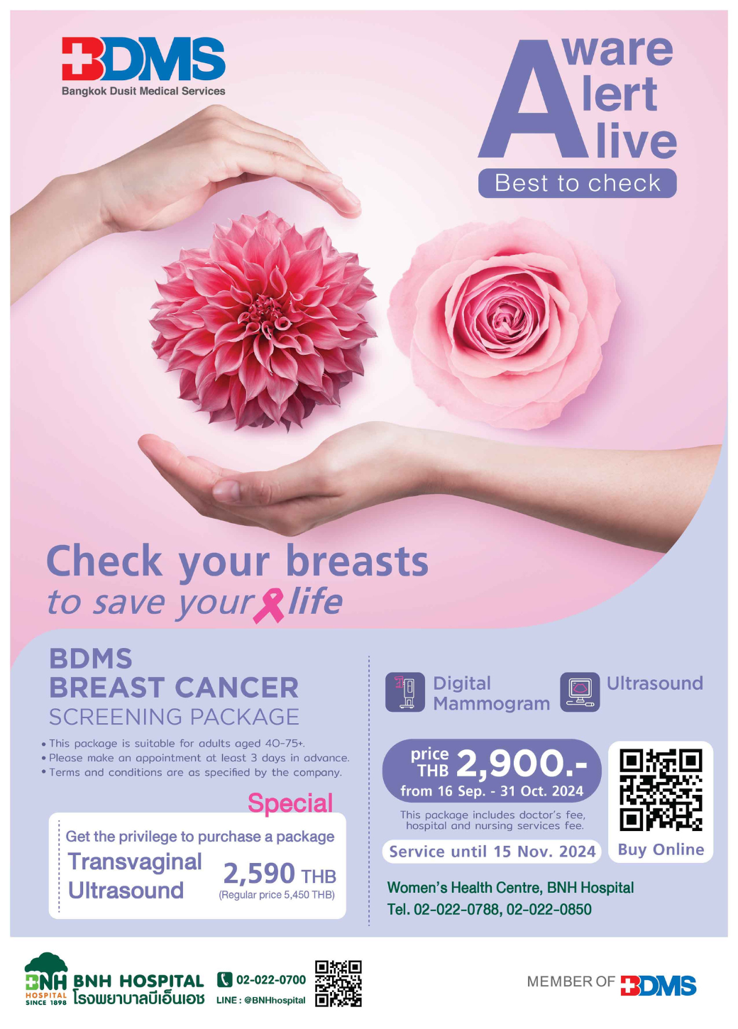
BDMS Breast cancer screening package
฿5,500
Breast Cancer: A Silent Threat You Shouldn’t Ignore
Here are some of the factors that can increase your risk of developing breast cancer:
- Age : Women over 50 are at a higher risk.
- Personal History of Breast Cancer : Individuals who have had breast cancer in one breast are at a 3-4 times higher risk of developing it in the other.
- Personal History of Ovarian Cancer : Due to the hormonal connection, women with ovarian cancer have an increased risk of breast cancer.
- Family History of Breast Cancer : Having a family member with breast cancer increases your risk.
- Exposure to Estrogen : Prolonged exposure to Estrogen, a female sex hormone, is linked to an increased risk of breast cancer.
- Lifestyle Factors : Obesity, lack of exercise, alcohol consumption, and exposure to high levels of radiation can increase the risk.
- Reproductive History : Women who have never had children or had their first child after age 30 are at a higher risk.
- Menstrual History : Women who started menstruating at a young age or went through menopause later in life have an increased risk.
Breast Cancer Screening Program
Digital Mammogram and Breast Ultrasound Package for women aged 40 and above.
Examination List:
- Physical Examination by a Specialist
- Ultrasound of Breasts
- Mammogram of Breasts
Terms and Conditions
- Price includes doctor’s fee, nursing and hospital services fee.
- Expenses for additional consultation, investigation, or treatment other than the package are not included.
- This package reserves the right for participating doctors only.
- This package is available until November 30, 2024 at Women’s Health Center on the 4th floor.
If you have no previous records at BNH, please register as a new patient at registration on the 1st floor. - Please make an appointment at least 3 days prior to using the services via LINE @BNHhospital or @Mbrace or call BNH Women’s Health Centre, 4th floor at 02-022-0788 Ext. 02-022-0850.
Mammogram is a diagnostic imaging procedure that uses low-energy X-rays to examine the breast tissue. It can be employed as a screening tool for asymptomatic individuals or to investigate specific symptoms. Studies have demonstrated that annual mammograms significantly enhance the detection of breast cancer in its incipient stages, thereby improving the likelihood of a complete cure and avoiding the necessity of a total mastectomy.
Digital Mammogram with Tomosynthesis
- 3D mammogram is a new type of mammogram that takes both 2D and 3D images of the breast at the same time. It provides a more detailed view of breast tissue compared to traditional mammograms.
- It is a machine that creates 3D images of the breast by capturing multiple images from different angles, then combining them to form a complete 3D image of the breast tissue.
- The resulting breast images are high-resolution, allowing radiologists to clearly see overlapping breast tissue and accurately pinpoint the location of any lumps. This provides a detailed diagnosis and can detect breast cancer at its earliest stages, significantly increasing the chances of a successful cure.
- A biopsy can be performed using a mammogram-guided needle to precisely target and remove tissue samples. This technique is highly accurate and particularly useful for examining calcifications that may not be visible with other imaging methods.
Indications for 3D Digital Mammography
- Screening for early-stage breast cancer: Used to detect breast cancer before any symptoms appear.
- Diagnostic tool for symptomatic patients: Used to diagnose breast conditions in women experiencing symptoms such as lumps, pain, or abnormal nipple discharge.
- Annual screening for women aged 40 and over: Recommended for women aged 40 and above to detect breast cancer early, increasing the chance of a cure and potentially preserving more breast tissue.
- Personalized screening for high-risk individuals: Women with a personal or family history of breast cancer should consult with a healthcare provider to determine the appropriate screening age and frequency.
Advantages of 3D Digital Mammography:
- Improved detection of small tumors: Mammograms allow doctors to detect smaller tumors, leading to more treatment options and a higher chance of a cure.
- No residual radiation: There is no remaining radiation in the body after the examination.
- Minimal radiation exposure: The amount of radiation used for diagnostic purposes typically does not have adverse effects on the body.

Should I have a breast exam before starting hormone therapy?
It is recommended to have a breast exam before starting hormone therapy, as many hormone treatments are contraindicated for individuals with breast cancer or those at risk. These medications can potentially cause cancer cells to spread and grow.
Can I take hormones or medication if I have a breast lump?
If a breast lump is diagnosed as benign or a cyst, hormone therapy may be an option. However, regular check-ups with a specialist are recommended to monitor the lump and receive appropriate guidance.
Should I get a breast exam before getting a breast augmentation?
Before breast augmentation, a breast examination by a specialist is recommended. This is because individuals diagnosed with or at risk of breast cancer should complete their treatment before surgery.
Are breast examinations still necessary after breast augmentation, and what is the appropriate screening method?
Individuals who have undergone breast augmentation with silicone or saline implants are advised to undergo regular breast examinations by a specialist. Both mammography and ultrasound can be safely performed on patients with implants. Mammograms for these individuals typically include an additional view showing the breast with the implant to assess the integrity of the implant, such as any leakage or fluid accumulation. An additional view, compressing only the breast tissue while displacing the implant, is also taken to evaluate the breast tissue for any lumps or calcifications.
Breast Self-Examination Guidelines
BNH Hospital recommends that women aged 40 and over have an annual breast exam.
Where can I get this service?
You can visit the Women’s Health Center on the 4th floor of BNH Hospital.
If you have no previous records at BNH, please register as a new patient at the Medical Records Department on the 1st floor.
Do I need to make an appointment?
To ensure a smooth service experience, it is advisable to make a reservation at least 3 days prior to your desired appointment time via Line.
Is it possible to schedule an appointment with my primary care physician?
This package reserves the right for participating doctors only.
Is this product/service transferable to another person?
This service/package is transferable to another person.
Is this covered by insurance?
The specific terms and conditions of your insurance policy will determine the procedures for obtaining a receipt. Please contact your insurer or agent for assistance.
Can I get a medical certificate?
A medical certificate confirming the visit can be requested.
Can I use the service after I buy it?
Walk-ins are welcome based on availability, however, we strongly recommend making a prior appointment for a more seamless experience.
Can I get a breast cancer screening even if I don't have any symptoms?
Even if you have no risk factors and no symptoms, you can still get screened.
- For women aged 35-40, an ultrasound is recommended.
- For women aged 40 and over, a mammogram is recommended every year until around age 70. If no abnormalities are found after age 70, screening can be done every other year.
- Women with a family history of breast cancer should start screening at age 35 or 10 years before the age at which their closest relative was diagnosed, whichever comes first. Close relatives include mothers, sisters, daughters.
Can I have a breast cancer screening even though I'm under 40?
Initially, we recommend a breast ultrasound. Mammography is most effective in detecting small breast cancers in women over 40 due to less dense breast tissue compared to younger women. Therefore, mammograms may be harder to interpret in younger women. However, if you have a family history of breast cancer, annual screenings should begin at age 35 or 10 years before the age at which your closest relative was diagnosed, whichever is earlier.
Is it possible to undergo an ultrasound examination solely, given that I am over 40 years of age and have no associated symptoms or medical history?
It is recommended to undergo an ultrasound examination.
Is a mammogram sufficient for a screening if I am over 40 with no symptoms or relevant medical history?
While mammograms provide detailed images, they cannot definitively distinguish between solid masses and fluid-filled cysts. Therefore, it’s essential to complement mammograms with ultrasound examinations. Mammography is highly effective in detecting calcifications and masses, while ultrasound helps differentiate between cysts and solid or malignant tumors. Additionally, ultrasound is crucial for confirming the absence of masses in women with dense breast tissue, often due to hormone replacement therapy or younger age.
General preparation for a medical examination
- The ideal time for a mammogram is 7-14 days after the end of your menstrual period.
- Avoid applying lotion, powder, deodorant, or any other topical products to the breast and underarm areas. These substances can mimic the appearance of calcium deposits often found in breast cancer on a mammogram, leading to misdiagnosis.
- Pregnant or potentially pregnant patients should inform the staff before the procedure. Mammograms are generally avoided during pregnancy, and an ultrasound will be performed instead.
What does it feel like to have a mammogram?
- During a mammogram, you will need to stand still while the machine compresses your breast and takes an image of each breast. This process takes about 5 seconds per breast. You may experience slight discomfort, which is necessary to obtain clear images and detailed views of your breast tissue. Typically, two standard views are taken of each breast. After the mammogram, you will undergo a breast ultrasound performed by a radiologist, which takes approximately 20 minutes.
- Individuals who have undergone breast augmentation with silicone or saline implants can safely undergo mammograms without any harm to the implants. During the imaging process, an additional view is taken compared to a standard mammogram. This extra view captures the breast with the implant and a separate view of the breast tissue alone by pulling the implant backward.
- The amount of radiation exposure from a mammogram is very low and only affects the body during the brief exposure to X-rays. It does not accumulate in the body and cannot be transmitted to others.
- Feeling relaxed and avoiding tension can make the examination easier and reduce discomfort from breast compression. Many patients are concerned about the discomfort associated with mammograms, but the benefits far outweigh the potential discomfort.
Can I have a mammogram while I'm pregnant?
Patients who are pregnant or suspect they may be pregnant should inform our staff beforehand. Mammograms are not recommended for pregnant women, and only ultrasounds will be performed.


Related products
Original price was: ฿16,300.฿13,000Current price is: ฿13,000.


Original price was: ฿4,020.฿1,690Current price is: ฿1,690.