Subdural Hemorrhage: Non-surgical Treatment, Safe, and no surgical incision
Chronic Subdural Hematoma (CSDH) is a significant medical condition commonly found, especially in the elderly. If left untreated, it can lead to severe consequences, including disability or death.
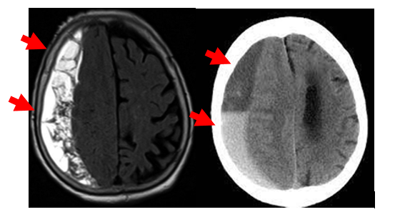
MRI and CT scan images of the brain showing right-sided CSDH (Chronic Subdural Hematoma) with compression of the brain (as indicated by the arrows).
Causes:
Head injury, even minor impacts
Use of blood-thinning medications
Regular alcohol consumption
Liver diseases affecting blood clotting
Brain atrophy in the elderly, increasing subdural space
Brain surgery or cerebrospinal fluid drainage
Symptoms of CSDH:
Headache: Most common symptom, can be continuous or intermittent
Weakness: May affect one side of the body
Numbness in various body parts
Slurred speech or communication problems
Seizures: Especially with large hematomas
Confusion: Disorientation about time, date, or place
Memory loss: Difficulty remembering recent events or short-term information
Impaired decision-making: Problems with analysis or decision-making
Unsteady gait or poor balance
Poor muscle coordination
Mood swings: Irritability or depression
Personality changes: Behavior different from usual
Importantly, these symptoms often develop slowly, making it difficult for patients or relatives to notice initially.
Treatments:
Observation (for mild cases with small hematomas)
Burr Hole Surgery: Small holes drilled in the skull to drain blood
Craniotomy: For large hematomas or recurrent bleeding
Disadvantages of surgery: Risks from anesthesia, potential complications like infection, longer recovery, and possible recurrence.
4. Endovascular Embolization
Treatment of chronic subdural hematoma by embolization is a new non-surgical option. It involves Middle Meningeal Artery (MMA) Embolization to reduce blood flow to the outer meninges, the source of bleeding in CSDH. Advantages include:
Safer than surgery
No surgical incision
Less painful for patients
Good treatment outcomes
Immediate resumption of blood thinners or antiplatelet medication if needed for other conditions
Treatment choice depends on factors like hematoma size, symptom severity, patient’s age and health, and available technology. Doctors choose the most appropriate method for each patient, focusing on the best outcome with minimal risk.
Treatment procedure:
Anesthesia or conscious sedation
Catheter insertion through the groin
Guiding the catheter to the affected meningeal artery
Injecting special embolization material through a microcatheter
The procedure takes about 1-2 hours
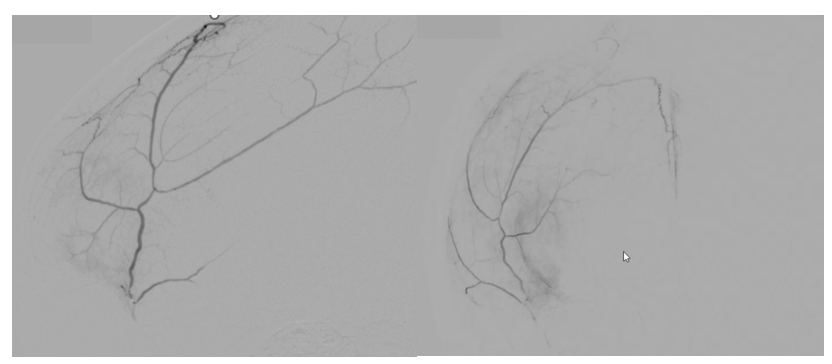
The image shows contrast injection into the Middle Meningeal Artery (MMA) supplying the meninges.
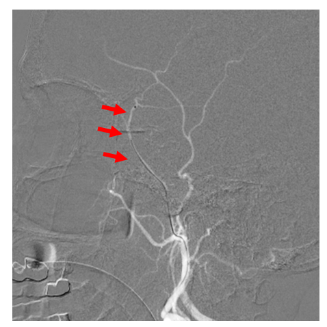
The arrow in the image shows the insertion of a microcatheter into the MMA (Middle Meningeal Artery), followed by embolization under DSA (Digital Subtraction Angiography) guidance.
Treatment outcomes:
Most patients improve within 1-2 weeks
Accumulated blood is gradually absorbed naturally
Follow-up:
Brain CT scans at 1 and 3 months post-treatment
Doctors assess hematoma resolution and patient symptoms
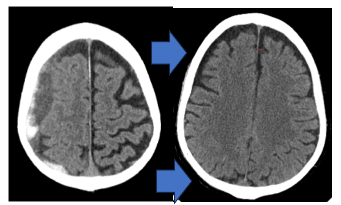
CT scan images comparing before and after treatment of right-sided CSDH, demonstrating complete resolution without surgery
In conclusion
Treating CSDH with embolization offers a more efficient, safer, and patient-friendly alternative compared to traditional surgery. This represents a significant advancement in the treatment of neurological disorders. However, the choice of treatment method should be considered on an individual basis.
References:
Link TW, et al. Middle meningeal artery embolization for chronic subdural hematoma: Endovascular technique and radiographic findings. Interv Neuroradiol. 2018;24(4):455-462.
Ban SP, et al. Middle meningeal artery embolization for chronic subdural hematoma. Radiology. 2018;286(3):992-999.
*All copyright of pictures: my own photo (Dr. Ittichai Sakarunchai)
Written by
Assoc. Prof. Ittichai Sakarunchai, M.D.
Assistant Professor of Neurosurgery & Interventional Neuroradiology
BNH Neuroscience Centre
We use cookies to improve performance. and good experience in using your website You can study the details at Cookies policy and you can change your cookie settings by clicking Cookie settings
Are very essential to our website as these allow the web servers to respond to your actions, support the structure of the page being displayed to you as well as provide the consistency of experience during your journey of surfing our website. These cookies will last only till the end of your visit and will be deleted automatically since then.
See list of strictly necessary cookies
Analytical / Performance cookies
Allow us to monitor the performance of our websites by determining the number of page views and the number of unique users a website has. Web analytics services are performed so as to analyse patterns of user behaviour and we use that information to enhance user experience or identify areas of the website which may require improvement. Nonetheless, the information is anonymous (i.e. it cannot be used to identify you and does not contain personal information such as your name and email address) and it is only used for statistical purposes.
See list of analytic / performance cookies
Functional cookies
Help us to recognise when you revisit our website. With this information, thereby we can customise our website based on your preferences, provide enhanced, more personal features to facilitate your visit. Such information collected by these cookies is usually anonymised, hence we cannot identify you personally.
See list of functional cookies
Behavioural Advertising / Targeting cookies
Will be placed on your device to record your visit, the page and the link you have visited and followed. The gained information will be used to tailor our website and our advertising campaign to suit your interests.
See list of marketing cookies